Sinus Graft

The maxillary sinus is a hollow cavity, lined by a thin blanket (Schneiderian membrane), located on both sides of the upper jaw. It’s purpose is to: provide humidification of air, serve as a ” crumple zone” in trauma, and enhance vocal resonance. When the posterior teeth are extracted, the sinus “pneumatizes” or expands, decreasing the available height of bone. A sinus graft procedure augments the vertical height of bone for dental implant placement. This made be done before or in conjunction with dental implant placement.
Internal (Osteotome) Sinus Lift
The internal or osteotome sinus lift is performed usually at the time of implant placement. A minimum of 3-5 mm of vertical bone height is recommended. The bone is prepared to just beneath the floor of the sinus, which is then gently tapped/infractured. Bone graft is introduced through the osteotomy (drilled hole), lifting the Schneiderian membrane from the walls of the sinus. An implant fixture can be placed simultaneously.
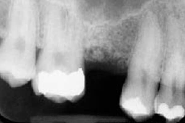
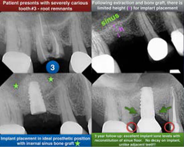
Figure 1: Pre-surgical
This patient has approximately 6-7mm of vertical bone beneath the floor of the sinus.
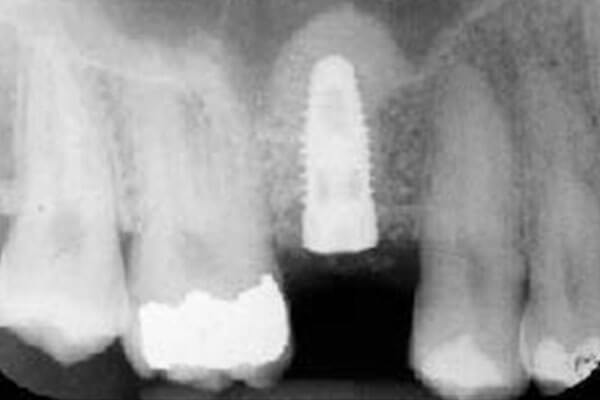
Figure 2: Post surgical
An internal sinus graft was completed and the implant placed simultaneously. Note the domeshaped elevation (bone graft), indicating excellent graft containment.
Lateral Window (Modified Caldwell Luc) Sinus Lift
In severely resorbed/atrophic ridges, there is inadequate vertical bone remaining, requiring more bone grafting into the sinus. A small window is prepared on the side of the maxillary sinus, and the Schneiderian membrane is gently elevated from the surrounding walls. Bone graft is placed and the window is covered with a barrier membrane and the tissue sutured. A 6-9 month maturation period is required prior to implant surgery.
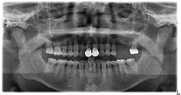
Figure 1:
Patient presents with severely vertically deficient bone upper left posterior. The panoramic image shows that there is 2mm of residual bone height, inadequate for implant placement. Lateral sinus grafting is necessary to create vertical height for implant placement.
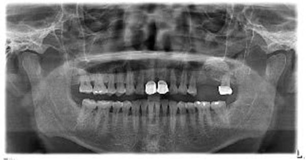
Figure 2:
6 months following sinus grafting, increased vertical bone height is noted as well as increased graft mineralization (upper left segment).
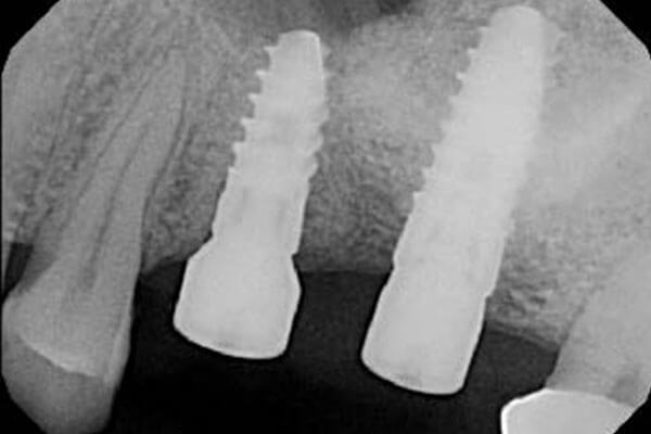
Figure 3:
An x-ray taken after implant surgery shows the final position of the dental implant relative to the newly formed sinus graft. The bone was completely mineralized, allowing the implant to be inserted “tightly” for stability.
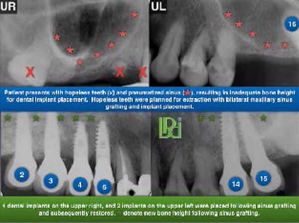
Additional Cases: