Cone Beam Scanner
Our office utilizes the latest technology, incorporating 3 dimensional cone beam scanning, offering more information for proper planning of advanced bone grafting and implant surgeries (ig. precise nerve location, sinus anatomy, bone volume, etc.).
Traditional 2 Dimensional Radiographs
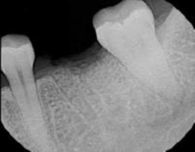
Digital radiographs show incredible detail and minimize radiation exposure.
However, the exact location of the mandibular nerve is not very clear in this radiograph.
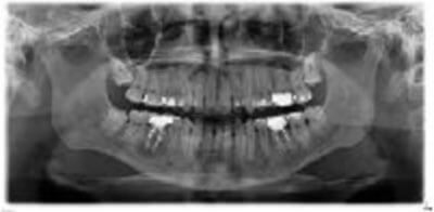
A digital panorex is often utilized in conjunction with traditional dental radiographs, providing more information on anatomy – sinus, mandibular nerve, location of wisdom teeth, etc.
However, we are unable to visualize depth (or thickness) of bone, lesions, exact positions of teeth.
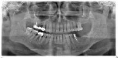
The top right bridge has failed and was slated for extraction. A lateral window sinus graft was performed and allowed to heal for 6 mos.
3 Dimensional Imaging
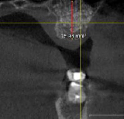
Following healing, a 3D scan was taken to evaluate: bone height (red line) as well as bone thickness. Evaluation shows adequate bone volume to place an implant fixture.
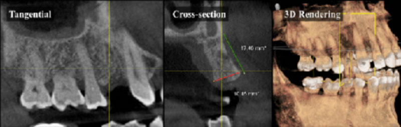
3D conebeam scan of a proposed implant site gives the surgeon more information in evaluating the site.
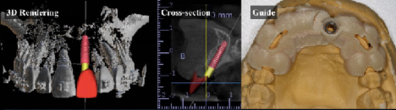
Using implant software, surgical placement of implants can be planned in ideal prosthetic position, and placement facilitated with a custom drill guide for greater precision and shortened surgical times.